PERSONALIZED WOUND HEALING
THE PATCH THAT USES YOUR BLOOD – TO HEAL YOUR WOUND
5 Reasons why 3C Patch® is leading the race in Advanced Wound Care

100% Autologous

Entirely alive

Remarkably simple
Immediately available

Completely proven
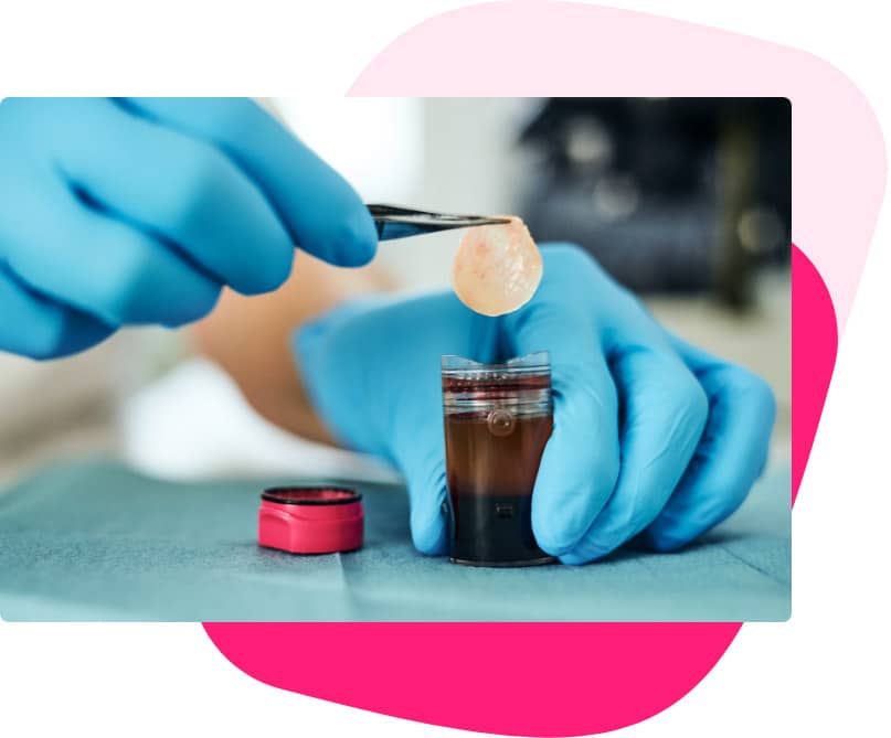
PERSONAL WOUND HEALING
The only 100% autologous wound care patch available
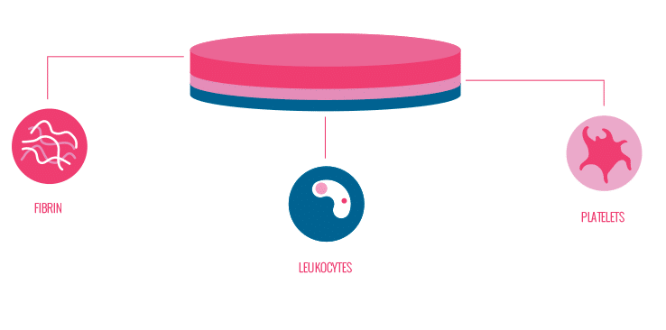
The proprietary centrifugation process creates a robust 3-layer patch containing fibrin, platelets, and leukocytes.

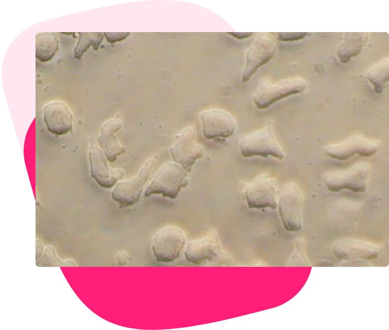
98% OF THE PLATELETS ARE RECOVERED
Entirely alive
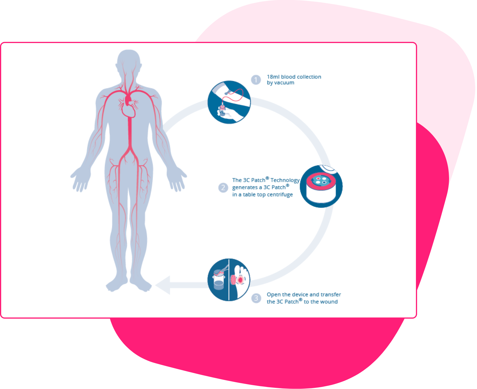
READY IN ONLY 20 MINUTES
Remarkably simple

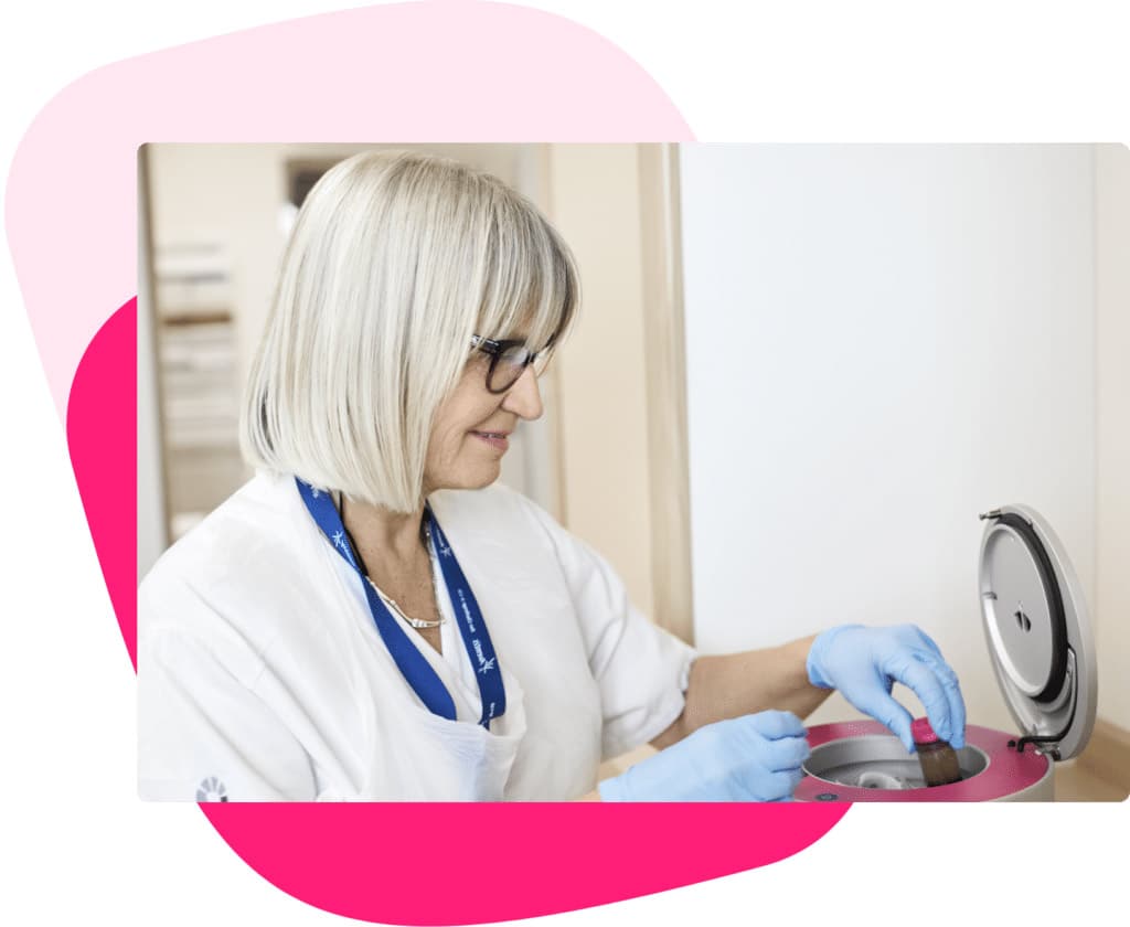
ON-SITE AUTOMATED PROCESS
Immediately available
3C Patch® is created in 15-20 minutes through a completely automated centrifugation process, at the end of which the 3C Patch® is ready to be transferred onto the patient’s wound.
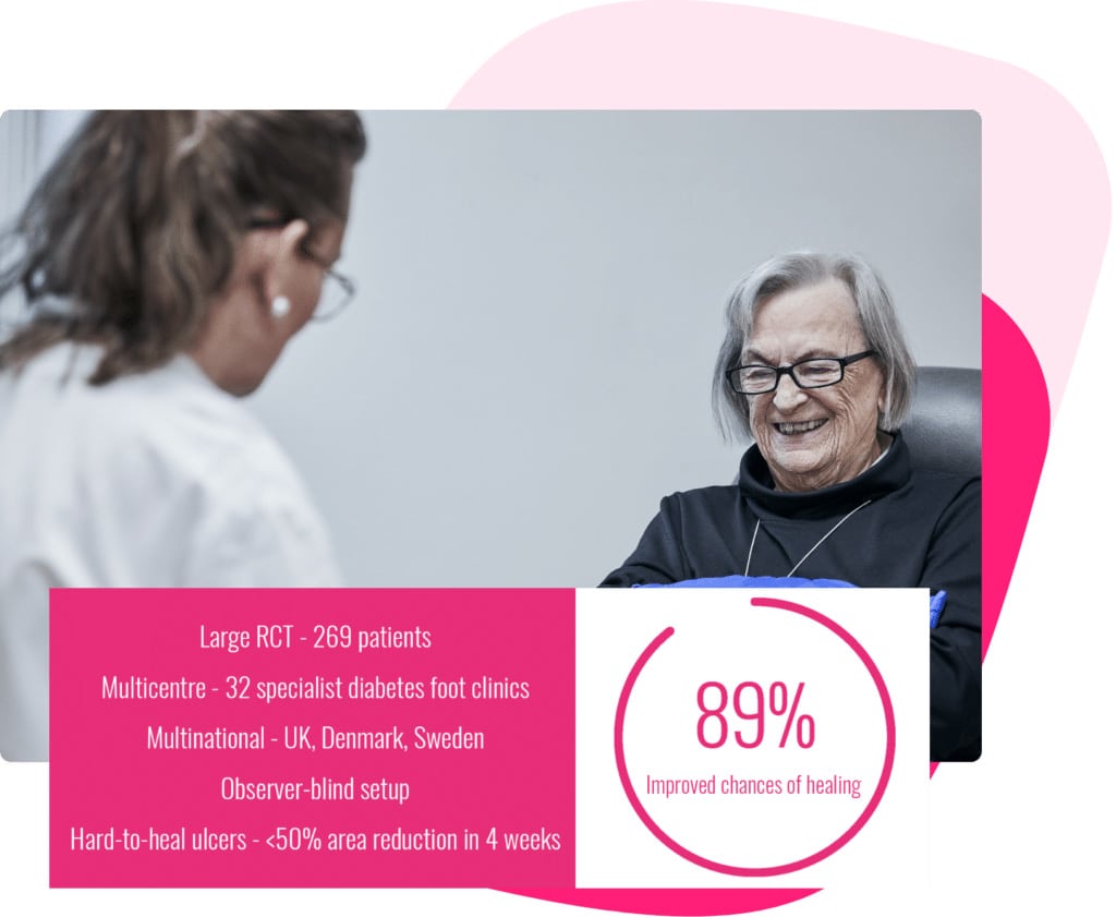
COMPLETELY PROVEN
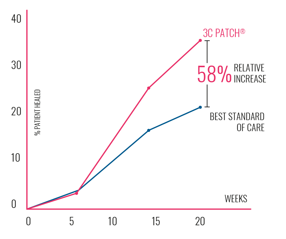
Significantly increasing the chances of healing
Read more about the clinical data that supports the effectiveness of 3C Patch as an advanced wound management solution.

3C Patch® Mode of Action
Watch the video explaining everything
WANT TO KNOW HOW IT WORKS IN REAL LIFE?
3C Patch® is not a wound care dressing – it’s an advanced wound care procedure done by the provider at the point-of-care
By comparison, using a 3C Patch® is like getting “a blood transfusion for your wound.” All the healing components from the blood are reformulated through an automatic centrifugation process into a 3-layered patch containing approx. 50 million leukocytes known to fight infection, 3 billion platelets having high growth factor levels, and fibrin, which provides moisture retention, structural integrity and makes it easy to handle.
PATIENT COMPLIANCE
How do patients react to having 3C Patch® as their wound care treatment
Furthermore, the randomized controlled trial results reported no device-related serious events, no differences in adverse events or serious adverse events between the control group and the 3C Patch® group, and no differences in new anemia between groups, despite weekly blood sampling in the 3C Patch group.
What are the main criteria for adding a new advanced wound care product in your clinic?
The science behind the product
The validity of the clinical data
Availability and implementation
Cost and reimbursement
Science and mode of action of 3C Patch®
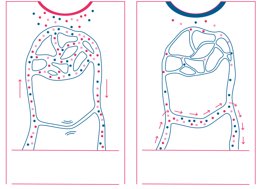
In people with diabetes, continuous high blood glucose levels lead to neuropathy. This results in non-functional AV-shunting and impaired microvascular flow, which causes chronic capillary ischemia. When the AV shunts become denervated, they lose their contraction response and stay open. This stalls the inflammatory response – which is key to healing – and prevents a sufficient number of immune cells, platelets, growth factors, and other signaling substances necessary for healing from reaching the wound.
Applied directly to the wound surface, 3C Patch® provides a high concentration of fibrin, leukocytes, and platelets. Fibrin provides moisture retention; leukocytes are known to fight infection, while platelets are known to provide high growth factor levels and participate in the resolution of inflammation.
Clinical data
3C Patch RCT Study design:
• An independent multicenter, multinational, observer-blind, investigator-driven Randomized Controlled Trial on patients with hard-to-heal Wagner grade 1-3 DFUs
Intervention:
• 3C Patch® was applied once a week for up to 20 treatments, or until the target ulcer was completely epithelized.
In the first treatment session, the ulcer was sharp debrided before one or two patches were transferred to the ulcer, which was then covered by a primary dressing. Secondary bandages were applied as decided on a case-by-case basis and changed depending on exudate levels
Patients:
• 269 patients were randomized
• “Hard-to-heal” defined rigorously as having DFUs that did not reduce in area more than 50% over a 4-week run-in period despite best standard of care according to IGWDF (incl. ref) guidelines 2019 including debridement, offloading etc. as appropriate
• The groups were well matched at baseline and consisted of those in most need of new treatment, those with “hard-to-heal” DFUs
|
Evidence from the large randomized controlled trial confirms strong clinical efficacy on chronic diabetic foot ulcers, compared to the best standard of care. Read more |
Availability and implementation
3C Patch® is immediately available at the point of care. To create a 3C Patch® you need to follow the procedure described in the next steps:
1. 18 ml of the patient’s blood is drawn into a 3C Patch® Device by venipuncture
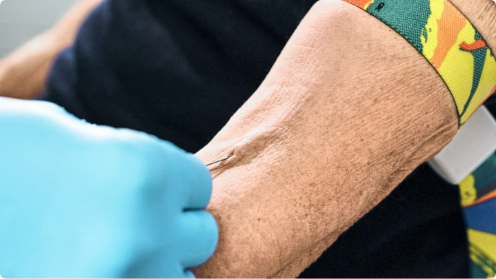
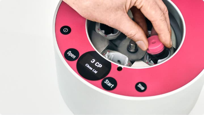
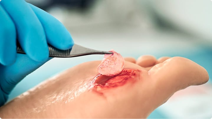
Easy to use and fits well within the clinic’s workflow
Cost and reimbursement
FAQ
Frequently asked questions
How do I prepare the ulcer before applying the 3C Patch®?
How many 3C Patch® treatments are needed?
Can I treat deep ulcers, interdigital ulcers, or ulcers with exposed tendons/bone contact?
Can I cut the 3C Patch® into shape so it fits the ulcer?
[2] Game F., Jeffcoate W., Tarnow L., Jacobsen JL., Whitham DJ., Harrison EF., Ellender SJ., Fitzsimmons D., Löndahl M., LeucoPatch system for the management of hard-to-heal diabetic foot ulcers in the UK, Denmark, and Sweden: an observer-masked randomized controlled trial. Lancet Diabetes Endocrinol. 2018;6(11):870-878.
[3] Lundquist, Jørgensen, Fagher, Löndahl (2015). 3C Patch™ – an adaptive triple layered autologous platelet and leukocyte rich fibrin patch and its clinical use in the treatment of hard-to-heal diabetic foot ulcers. Poster, Innovations in Wound Healing 2015.
